Most common cardiac mass ➜ thrombus
Metastasis: 20-40 times more frequent than primary tumor
– from lung and breast
Primary cardiac tumor: prevalence 0.002-0.3%
Benign: 75% of primary cardiac tumor
– Most common : myxoma
Malignant: 25% of primary cardiac tumors
– Most common : angiosarcoma
Pericardial tumors
– benign teratomas, malignant mesotheliomas
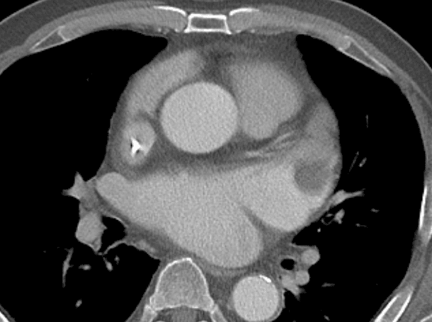
According to JACC 2010 criteria CT evaluation of cardiac mass is inappropriate
➜ If suspicious, get cardiac MRI


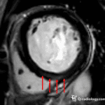
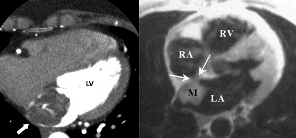
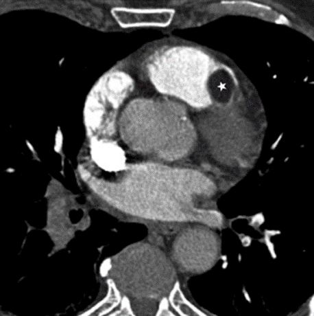
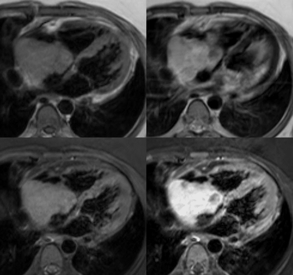
1. Left atrial myxoma
– A polypoid, intracavitary
– Left atrial mass (75%) from the interatrial septum (around fossa ovalis)
– Narrow base with stalk
– Calcification
– Size : 4~8 cm
– Cause of embolism, about 35% LA myxoma are related to embolism
– Prevalent age : 20~50
– MRI finding
– iso signal intensity to myocardium on T1WI
– high signal intensity or low signal intensity on T2WI (due to hemosiderin or calcifications)
– heterogeneous enhancement
– Treatment : Surgery
– Differential diagnosis : thrombus


2. Lipoma
– Male = Female
– Adult > Children
– Size : 1~15 cm
– Any location in the cavity, myocardium, epicardium
– Fat containing mass with no enhancement
– Differential diagnosis : Lipomatous hypertrophy of interatrial septum


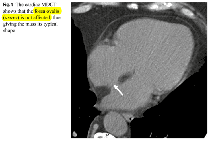
2-1) Lipomatous hypertrophy of interatrial septum
– Elderly, obese individual
– Characteristic dumbbell shape, with relative sparing of the fossa ovalis

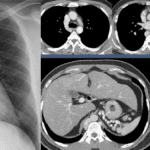
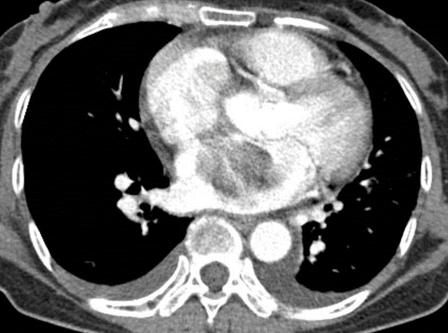
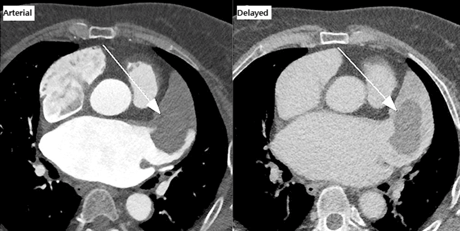
3. Primary cardiac angiosarcoma
– Most common malignant cardiac tumor
– Male > Female ; rare in children
– Prevalent location is the right atrium; frequently invades pericardium
– Symptoms
– Right heart failure, cardiac tamponade
– Frequently accompany with pericardial effusion
– No effective treatment
– Imaging finding
– Iso-signal, nodular lesion in both T2WI and T1WI (“cauliflower” appearance)
– Linear enhancement with diffuse pericardial invasion (“sunray” appearance)

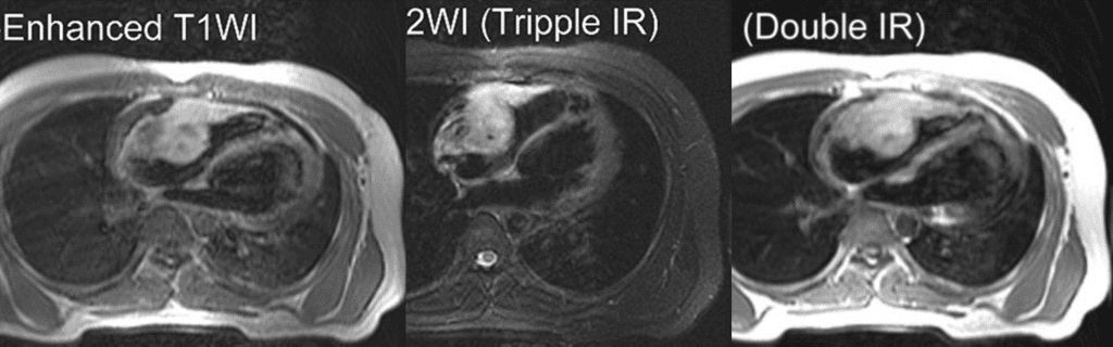


Tissue within the pericardial cavity contained non-enhancing regions separated by enhancing lines radiating from epicardium to pericardium, this pattern of enhancement (previously described as “sun ray appearance”)
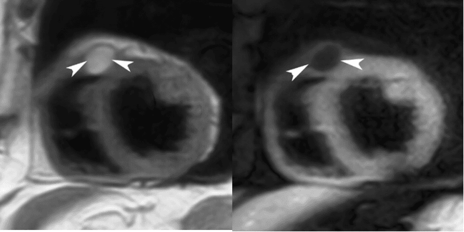
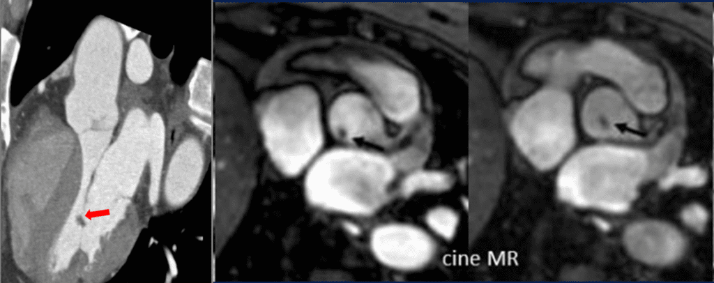
4. Papillary fibroelastoma
– Most common tumor of valve
; Aortic valve > Mitral valve > mitral chordae tendinae, RA endocardium, endocardial surface of ventricles
– Less than 1.5cm, short thin stalk
– Potential source of systemic emboli
– Differential diagnosis : Vegetation, thrombus

5. Cardiac thrombus
– Most common intracardiac mass
– Cardio-embolic stroke : 10-20%
– Risk factor
; Atrial fibrillation, valve disease, severe LV dysfunction
– Location
; most common in Left atrium or Left atrial appendage
; LV – severe in LV dysfunction
– Differential diagnosis
; tumor (myxoma), circulatory stasis


5-1) Differential diagnosis : Tumor vs. Thrombus


5-2) Differential diagnosis : Circulatory stasis